
Autonomous Microscope
It is estimated that Microalgae produce up to 50% of the oxygen we breathe, making them a valuable resource to all terrestrial species including ourselves. Additionally, as one out of every seven people on Earth depends on seafood for their daily protein requirements, understanding the microalgae at the root of the marine food web is important to a large number of people's survival. In recent years, it has been proposed that shifts in planktonic populations and species diversity correlate to the rise of water temperatures and acceleration of marine climate, pointing to the possibility of a large impact on the entire marine biome. This is why marine scientists worldwide are making these tiny organisms the focus of their research.
The analysis of the ocean surface water communities is at the heart of the marine scientists' work. Traditionally, this process required collecting in-person samples during expensive and time intensive boat expeditions to evaluate later. Jupiter Research Foundation developed a compound microscope system that travelled on an autonomous, unmanned Wave Glider and allowed in-situ water sampling, immediately screening for microorganisms or other particles. The microscope then took date/time/geolocation stamped pictures of anything it found and sent these pictures back to the lab in near real-time.
Not only was this approach significantly less expensive than the traditional sampling process, providing budget for a larger number of autonomous expeditions, it also provided a greater spatial temporal range of sampling to produce better informed predictions of future microalgae patterns.
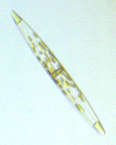
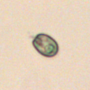
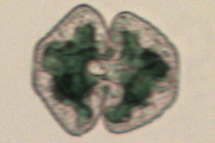
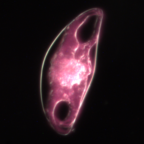
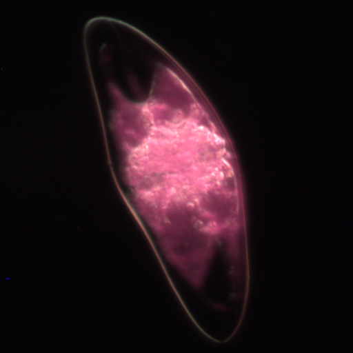
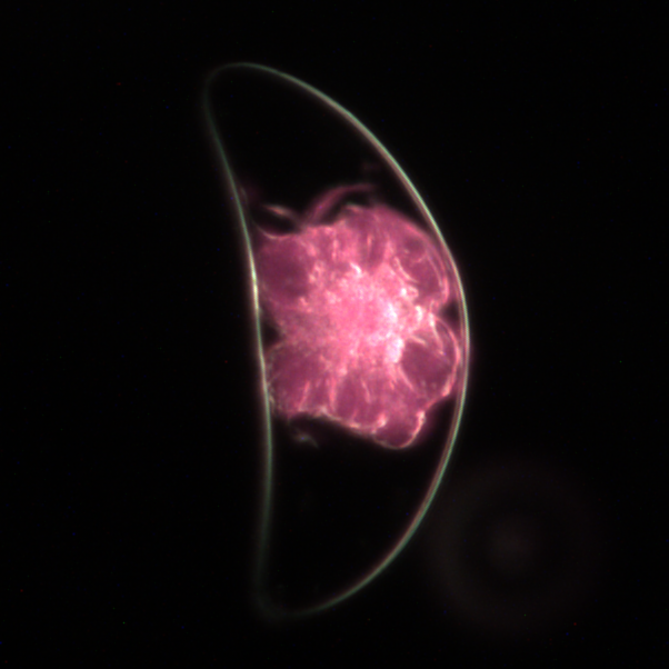
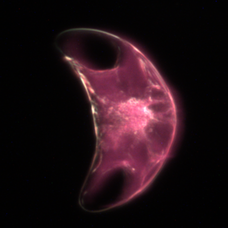
Another unique feature of our Autonomous Microscope was its combined use of darkfield microscopy and PS2 Fluorescence to produce both striking and recognizable images. Although the microscope prototype originally utilized brightfield, it was found that darkfield imaging ultimately aids in the distinguishment of phytoplankton and zooplankton and yields optical concentration of samples by allowing multiple exposures per image. Darkfield microscopy illuminate the membranes or edges of an object against a black background by configuring the condenser to pass only the light scattered by the specimen through the lens objective. This is opposed to brightfield microscopy where light is directed by the condenser through the specimen and then continues through the lens objective. The PS2 chlorophyll reaction takes place when a specific wavelength of blue light is focused down on the specimen, stimulating the species' chloroplasts to flouresce in a red wavelength then captured by the microscope's camera. What results are images of starkly outlined organismal membranes enclosing red organelles.

Autonomous Microscope payload getting ready for Canon 2015
Jupiter’s microscope team attempted to produce solutions to other problems and inquiries that arose when dealing with an autonomous plankton sampler. These included biofouling of the lens, imaging of larger and smaller specimens, and the creation of a photo library to automatically identify recorded organisms.